What is Corneal Topography?
Corneal topography, also known as corneal topometry, is a diagnostic method that is computer-assisted and provides a 3D (three-dimensional) map of the surface curvature of the cornea. The cornea, often known as the front glass of the eye, is accountable for around 70% of the eye’s overall focusing power.
If the cornea is uniformly rounded, then the eye will have normal vision. However, if the cornea is excessively flat, too steep, or unevenly curved, then the eye will not have normal vision.
Corneal topography generates a comprehensive and visually appealing representation of the structure and power of the cornea. Your eye doctor will be able to obtain highly specific information regarding the state of the corneal surface using this method of analysis.
These particulars are utilized in the processes of diagnosing, monitoring, and treating a variety of eye ailments. In addition to this, contact lens fittings and surgical procedures, such as laser eye surgery, require their use for accurate planning.
In laser vision correction, the corneal topography map is utilized in conjunction with other tests to establish exactly how much corneal tissue will need to be removed to correct vision and what pattern of ablation will be employed. This data is then used to plan the procedure.
Diseases and injuries that can be healed with computerized corneal topography include
- Corneal Infections
- Abrasions of the cornea
- Deformities of the corneal surface
- Dry eye syndrome
- Transplantation of the cornea
- Scarring or opacities on the cornea
- Astigmatism that is irregular as a result of corneal transplants
- Corneal Topography Procedure
The corneal topography apparatus comprises a computer that is connected to a bowl of light that has a pattern of rings in it. The patient is required to sit in front of the bowl with his or her head placed against a bar for the duration of the diagnostic test. During this time, a number of data points will be generated.
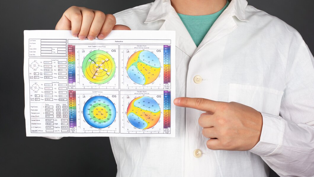
These data points are then digitized by computer software in order to make a printout of the shape of the cornea, with different colors being used to identify different elevations.
It is non-contact testing quick and does not cause any discomfort. Corneal topography gives medical professionals, specifically ophthalmologists, access to the most specific and in-depth information available regarding the curvature of the cornea, as well as potential vision problems and eye disorders.
Corneal topography is considered one of the best Contoura Vision Eye Surgeries in Delhi, as the majority of traditional testing methods fail to discover deformities of the cornea, such as keratoconus (a disorder that causes visual disruptions and impairments).
Patients opting for refractive surgery performed with an excimer laser undergo corneal topography. It is performed both before and after refractive surgery. This examination is considered to be of main relevance.
The corneal topography of an eye is utilized as a monitoring tool for the evolution of ocular surface problems such as pterygium, which is characterized by the growth of pinkish, triangular tissue on the cornea.
After undergoing corneal transplantation, a patient may experience irregular astigmatism, which refers to an imperfection in the cornea’s form.
Steps Involved in Performing Corneal Topography
Corneal topography of an eye is done to generate a map of the cornea, which provides the physician with the ability to develop a three-dimensional view of the shape of the cornea.
In corneal topography, a computer is typically connected to an illuminated bowl that contains a pattern of concentric rings.
In most cases, the patient will be instructed to sit in front of the bowl with a bar resting against the back of their head.
After that, multiple light concentric rings are projected onto the cornea, and the charge-coupled device (CCD) camera is used to capture the image that is reflected from the cornea.
After that, the data points are digitized by the software on the computer, and the corneal shape is printed out using a variety of color representations to indicate the varying elevations.
Cooler colors, such as blue and green, are used in the process as it becomes easier to locate portions of the cornea that are flatter. However, in some cases, more treacherous terrain may be indicated by warmer colors like orange and red.
Why is it vital to have a corneal topography?
Corneal topography, also known as computer-assisted video keratography (CAVK) or corneal mapping, is a technique of computer-assisted diagnostic imaging that helps create visuals of the corneal surface. This allows the eye specialist to view the visuals and analyze the entire shape of the cornea to diagnose and treat the patient.
It is a medical imaging method that does not include invasive procedures and is used to map the surface curvature of the cornea.
The topography of the cornea is of crucial relevance in defining the quality of vision because the cornea is generally responsible for around 70% of the eye’s refractive power. It plays a crucial role in the diagnosis process.
Wrapping up…
So, if you’re looking for a reputable place for corneal topography, look no further. Contoura Vision Global is one of the best places for Contoura Vision Eye Surgery in Delhi.
We have the most qualified experts on our team; we are able to provide you with the most accurate corneal topography and, based on that, the most accurate refractive surgery.